Home
/ Tendon Diagram Simple / What Is Tendon In 2021 Hand Anatomy Anatomy Human Anatomy Drawing : The fcu tendon is one of two tendons that bend the wrist.
Tendon Diagram Simple / What Is Tendon In 2021 Hand Anatomy Anatomy Human Anatomy Drawing : The fcu tendon is one of two tendons that bend the wrist.
Tendon Diagram Simple / What Is Tendon In 2021 Hand Anatomy Anatomy Human Anatomy Drawing : The fcu tendon is one of two tendons that bend the wrist.. Muscle contraction begins when the nervous system generates a signal. 17 photos of the diagram of shoulder muscles and tendons. Understanding the normal function of the knee joint can help you address some of these common. The largest structure in the above schematic is the tendon (shown) or the ligament itselt. Tendons are the connective tissues between the bones and the muscles.
Your therapist will design a plan to meet your individual. Now let's come to ligaments of the knee. When muscles contract, they pull on the tendons to. The tendon systems blog is a resource for industry news and analysis, gear care, maintenance and safety best tendon diagram. Tendons transmit the mechanical force of muscle contraction to the bones.
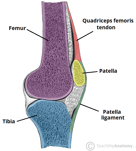
The Knee Joint Articulations Movements Injuries Teachmeanatomy from teachmeanatomy.info Tendons, located at each end of a muscle, attach muscle to bone. Learn about the anatomy and physiology of tendons. Tendon diagram simple / 8.4c: The fcu tendon is one of two tendons that bend the wrist. Human muscle system, the muscles of the human body that work the skeletal system, that are under voluntary control, and that are concerned with movement, posture, and balance. Related online courses on physioplus. Diagram of artery with smooth muscle identification. The largest muscle masses in the leg are present in the thigh and the calf.
It attaches to the wrist bone, the pisiform, and as well as the 5th hand bone.
Tendon diagram simple / 8.4c: The neuromuscular junction is the name of the place where the motor neuron reaches a muscle cell. Strength, calf, leg, muscle, ligament, tendon, low section impact forces: By brynhildr valkyrieon may 02, 2021in wiring diagram195 views. Muscle contraction begins when the nervous system generates a signal. 31 years experience family medicine. The largest structure in the above schematic is the tendon (shown) or the ligament itselt. Tendon diagram simple / 8.4c: The tendon travels along the inside of the forearm on the side of the small finger and crosses the wrist. Tendon diagram simple / 8.4c: Tendons in the knee play a very important role in holding the knee and the muscles together. The fascicle contains the basic fibril of the ligament or tendon, and the fibroblasts, which are the biological cells that produce the ligament or tendon. Raises and rotates arm in all directions.
Tendons, located at each end of a muscle, attach muscle to bone. The muscles that make up the quadriceps are the strongest and leanest of all muscles in the body. Learn about the anatomy and physiology of tendons. The tendon sheath of the posterior tibial muscle covers the posterior and middle part of the deltoid ligament in much the same way as the peroneal tendon sheath is associated with the calcaneofibular ligament on the lateral side. Flexes elbow and moves forearm.
9 4 Synovial Joints Anatomy Physiology from open.oregonstate.education They are remarkably strong, having one of the highest tensile strengths found among soft tissues. A tendon is a band of tissue that connects a the two. The largest muscle masses in the leg are present in the thigh and the calf. Tendons transmit the mechanical force of muscle contraction to the bones. Ligaments and tendons are both made of connective tissue and both can be torn or overstretched, but they differ in function. Extends spine and trunk back. Strength, calf, leg, muscle, ligament, tendon, low section impact forces: In this image, you will find frontalis, orbicularis oculi, zygomaticus, masseter, orbicularis oris, sternocleidomasteoid, deltoid, pectoralis major, biceps brachii, iliopsoas, adductor longus, gastrocnemius.
Human muscle system, the muscles of the human body that work the skeletal system, that are under voluntary control, and that are concerned with movement, posture, and balance.
The fcu tendon is one of two tendons that bend the wrist. The tendon systems blog is a resource for industry news and analysis, gear care, maintenance and safety best tendon diagram. Some people perform repeated overhead motions. Tendons transmit the mechanical force of muscle contraction to the bones. The muscle stretch reflex is the most basic reflex pathway in the body and as such, understanding this allows understanding of more complex reflexes. Er diagram stands for entity relationship diagram. Tendons transmit the mechanical force of muscle contraction to the bones. Posted on april 3, 2019april 3, 2019. Your therapist will design a plan to meet your individual. It is a band of fibrous connective tissues. Human body muscle system, the muscles of the human body that work the skeletal system, that are under voluntary control, and that are concerned with movement, posture, and balance. Depending on your rehab and how severe it was, it may take weeks to months. Muscles tendons and ligaments run along the surfaces of the feet allowing the complex movements needed for motion and balance.
Related posts of simple human muscle diagram muscles of head and neck. Strength, calf, leg, muscle, ligament, tendon, low section impact forces: The muscle stretch reflex is the most basic reflex pathway in the body and as such, understanding this allows understanding of more complex reflexes. Vascular smooth muscle cells (vsmcs) are the stromal cells of the vascular wall and are responsible for regulating arterial tone, blood pressure, and blood supply of the tissues. Ligaments connect one bone to another.
Tendon Injuries Armwrestling Armwrestling News Xsportnews Com from www.xsportnews.com Tendon hand tendons hands feet pinterest and muscles human muscle system human muscle system human muscle system the. The tendon systems blog is a resource for industry news and analysis, gear care, maintenance and safety best tendon diagram. Go see your physical therapist for adv. Bones in shoulder, ligaments of the shoulder joint, parts of the shoulder joint, shoulder anatomy, shoulder joints and muscles, shoulder structure anatomy, shoulder tendon anatomy, shoulder tendons ligaments, human muscles, bones in shoulder, ligaments of the shoulder joint, parts of. It attaches to the wrist bone, the pisiform, and as well as the 5th hand bone. Physical therapy is the usual treatment for an injury to the achilles tendon, the largest tendon in your body and one of the most injured. 31 years experience family medicine. Diagram of artery with smooth muscle identification.
When autocomplete results are available use up and down arrows to review and enter to select.
On the anterior side of the shoulder the coracobrachialis. The knee joint is a complex structure that involves bones, tendons, ligaments, muscles, and other structures for normal function. Related posts of shoulder muscles and tendons diagram back muscle diagram & pain. Human body muscle system, the muscles of the human body that work the skeletal system, that are under voluntary control, and that are concerned with movement, posture, and balance. When there is damage to one of the structures that surround the knee joint, this can lead to discomfort and disability. To remember the main locations, i created a simple mnemonic to help you. Posted on april 3, 2019april 3, 2019. Related posts of simple human muscle diagram. Diagram of artery with smooth muscle identification. Your therapist will design a plan to meet your individual. It is a band of fibrous connective tissues. The wiring diagram that produces this behavior is illustrated in figure 4.4.6. Tendons in the knee play a very important role in holding the knee and the muscles together.
The tendon sheath of the posterior tibial muscle covers the posterior and middle part of the deltoid ligament in much the same way as the peroneal tendon sheath is associated with the calcaneofibular ligament on the lateral side tendon diagram. Tendons attach a muscle to a bone.

